
Detecting cancer
Detecting cancer is a multi-stage process. Often, the patient will go to a doctor because of some symptom or other. Sometimes cancer is discovered by chance or from screening. The final cancer diagnosis is based on a pathologist’s opinion.
Different cancers are discovered in different ways
Detecting cancer, or cancer diagnosis, always entails detailed examination. Following a clinical examination by a doctor, a patient may undergo different methods of imaging and be referred for laboratory testing. The final cancer diagnosis is based on a pathologist’s opinion.
-
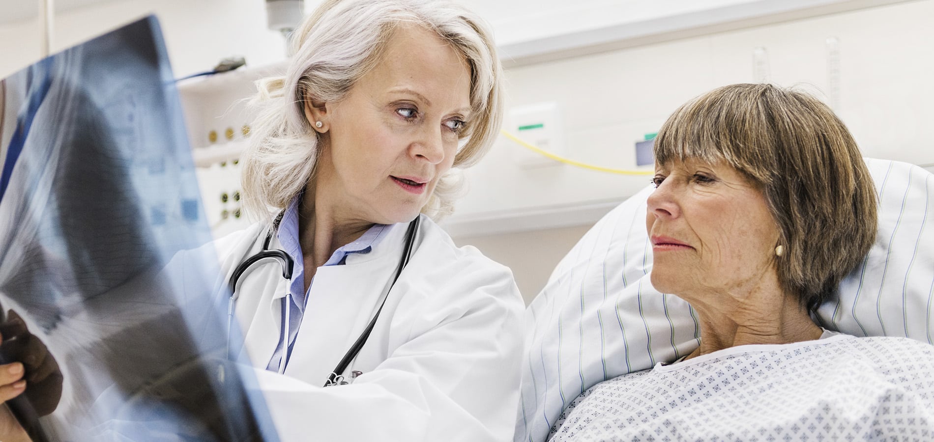
A general practitioner carries out a clinical examination of a patient. The doctor also considers the patient’s family background and charts the most common risk factors for cancer.
-
Cancer detection often involves radiological imaging. Imaging is also used to check the spread of cancer and progress of treatment, and to monitor cancer.
Oncological imaging is continually becoming more varied and accurate. Different imaging techniques aim to find the most suitable treatment option for each patient. Imaging techniques are often used in combination to obtain sufficient information.
The most common imaging method used to detect cancer and monitor its spread is Computed Tomography (CT), which provides cross-sectional imaging by computer. CT scans are made using X-rays.
Magnetic Resonance Imaging (MRI) is a procedure that uses powerful magnetic fields. This does not generate ionising radiation. Situations where MRI is used include examining cancer or sarcoma in the head and neck region.
Positron Emission Tomography (PET) is based on the faster metabolic rate of cancer cells compared to normal cells. With PET imaging the patient is given a radioactive tracer that is detected by scintigraphy. PET images can also be combined with CT.
Ultra sound examination is useful for examining the cervix, pancreas, liver and kidneys. Needle biopsies can also be taken in ultra sound examinations.
Endoscopic examinations are usually for inspecting the gastrointestinal tract, bronchial tubes, cervix, prostrate, bladder or head and neck region.
In mammography, an X-ray image is used to examine breast tumours. Mammography is also used in breast cancer screening.
In isotopic diagnostics a radioactive tracer is introduced into the patient’s body. The marker goes to the organ to be examined and various imaging methods can be used to determine whether the cancer has spread. Isotopic diagnostics can be used to identify the prevalence cancers such as breast, prostate and colorectal cancer.
-
Laboratory tests are carried out at the point when it is suspected that a patient has cancer. Normally, a blood sample is taken for monitoring blood counts.
With some cancers, determining cancer markers is useful. The number of tumour markers varies according to the cancer activity in the blood stream. Tumour markers can be used in cancer detection, monitoring and prognosis evaluation. The occurrence of markers can be established by a blood test.
Tumour markers are secreted by tumour tissue into the blood. Detecting tumour markers or their concentration may indicate the emergence of cancer or its recurrence. The sensitivity and accuracy of markers vary, and increase in their concentration does not always indicate the presence of cancer. They might not even be found in all cancer patients.
Examples of tumour markers include prostate-specific antigen (PSA) for prostate cancer and cancer antigen (CA) 15.3 for breast cancer.
-
Genetic research is talked about a lot in medicine these days. Genetic testing can be useful in connection with certain cases of cancer.
However, genetic testing cannot detect cancer, but can identify part of the genetic defects that indicate a predisposition to developing cancer, thereby detecting a genetic susceptibility to cancer. But so far there is no simple comprehensive genetic test for those interested in having a gene test.
-
A final diagnosis of cancer is based on an examination of tissue or cells under a microscope by a pathologist. Biopsies can be taken using a fine needle, large core needle or biopsy forceps, or the entire tumour may be removed by surgery.
A large core needle biopsy, or tissue sample, is almost always sufficient for confirming cancer diagnosis. The procedure involves removing a sample of a few millimetres from a tumour under local anaesthesia. The sample is examined to check if the tumour is malignant or benign, its gradation and distribution.
In addition or instead of a biopsy a cytological or cell sample can be taken. This can be done either as a fine needle biopsy or smear test.
A typical smear test is the Pap smear, used in gynaecology. Smear tests also include urine samples as well as samples taken from the gastrointestinal tract and cerebrospinal fluid.
A fine needle biopsy is taken with a thin needle or syringe. The accuracy of the sample depends on such things as the type of cancer. A fine needle biopsy can be taken from the thyroid gland, liver and lymph nodes. The malignancy of a tumour can usually be confirmed with a fine needle biopsy.
A cell sample result is given a five-class grading according to the classification of Pap test results, where 1 is benign and 5 clearly malignant.
Worrying about cancer-related issues?
The services of cancer organizations are intended for everyone who is wondering about issues related to cancer.